Fig. 1.

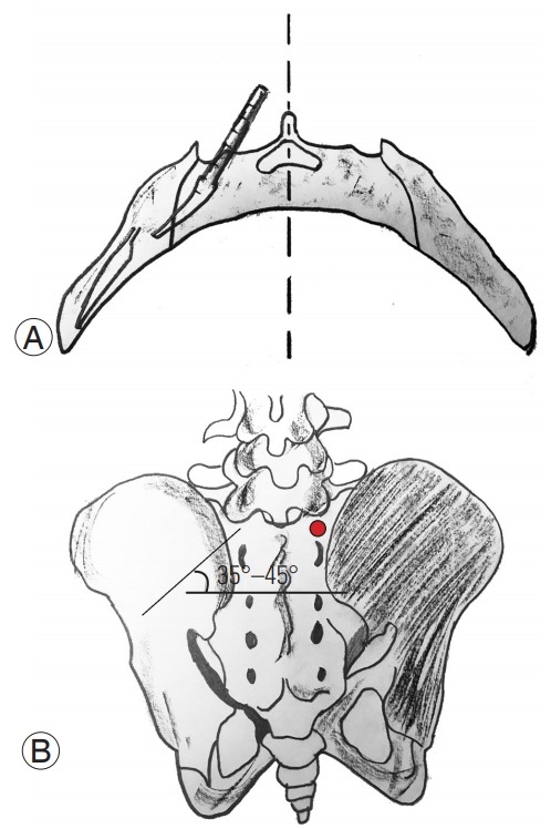
(A) Axial drawing showing the direction of the probe in the sacral and iliac bone. (B) Drawing showing the entry point (red dot) as well as the caudal direction of the screw (35°–45°).

(A) Axial drawing showing the direction of the probe in the sacral and iliac bone. (B) Drawing showing the entry point (red dot) as well as the caudal direction of the screw (35°–45°).