
Fig. 2.
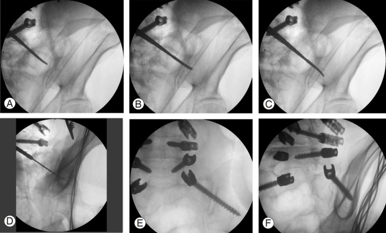
(A) Steps for S1 alar iliac implementation. The probe is against the sacroiliac joint. (B) Probe is advanced through the sacroiliac joint. (C) Probe is turned 180° and advanced in the iliac bone. (D) The ball-tipped feeler is used to detect the bony borders. A tear drop view is used for checking the final position. (E) Final position in the anteroposterior view. (F) Final position in the tear drop view.