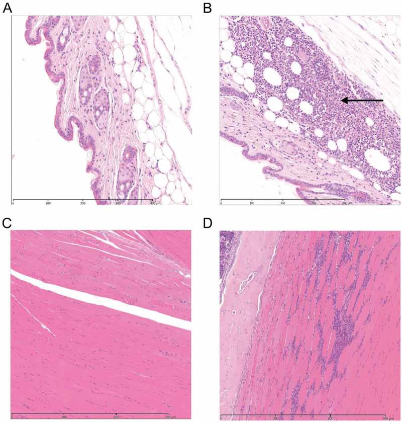
Figure 2.

Histological features in soft tissue with or without contusion.
Photomicrographs of H&E stained tissue sections 24 and 48 h following contusion. (representative of 12 per group). A. Control subcutaneous tissue at 24 h. B Focal inflammation in the subcutis of injured leg 24 hours after contusion. Arrow represents the area of injury with an inflammatory response. C. Control muscle tissue at 48 h D Injured muscle at 48 h after contusion with focal loss of myofibres and replacement by a mixed inflammatory response of neutrophils and macrophages.
