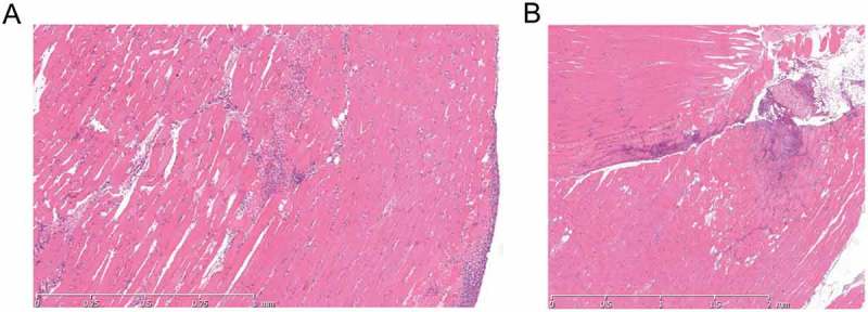
Figure 3.

Histological features in soft tissue are similar following infection without or with contusion.
H & E stained tissue sections following GAS thigh muscle infection. A Photomicrograph of GAS-infected thigh tissue 24 h after infection without contusion. B Photomicrograph of GAS-infected thigh tissue 24 h after contusion and GAS infection. Both sections show multifocal mixed inflammatory cell infiltrate with slight myodegeneration and cell lysis. Some haemorrhage is seen in A.
