
Table 3.
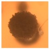
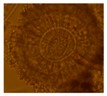
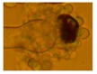
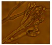
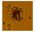
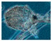
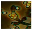
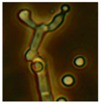
The eight fungal morphotypes isolated from settled dust collected before and after the ventilation improvement, characterized by toxigenicity, pathogenic potential, and conidiophore morphology.
| Toxicity | Colony Color | Size of Conidia/Spores | Morphology under Light Microscope | |||
|---|---|---|---|---|---|---|
| Growth at 37 °C | BSMI | ICP | MEA | (μm) | ||
|
Aspergillus section Nigri 1 strain |
+ | - | + | Black | 3.5–5 |
|
|
Asp. westerdijkiae 2 strains |
- | + | + | Yellow | 2.5–3 |
|
|
Eurotium sp. 1 strain |
+ | + | + | Green | 5–7 |
|
|
Penicillium sp. 10 strains (Terverticilliate) |
Green | 3.4 |
|
|||
|
Penicillium sp. 3 strains (Monoverticilliate) |
Green | 2.3 |
|
|||
|
Rhizopus sp. 10 strains |
- | - | - | Grey | 5–10 |
|
|
Trichoderma citrinoviride * 10 strains |
+ | + | + | Green | 1.6 × 3 |
|
|
Trichoderma sp. 5 strains |
- | + | + | Green | 4 |
|
* Identified to species level by ITS sequence analysis.