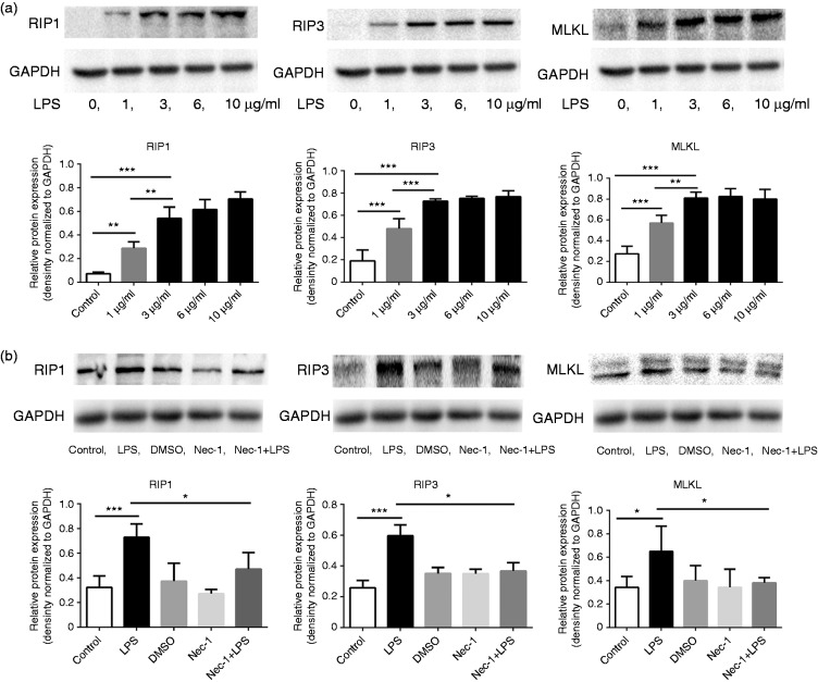
Figure 3.
Activated macrophages express upregulated RIP3 signaling. (a) RAW264.7 macrophages were exposed to varying concentrations of LPS (0 µg/ml, 1 µg/ml, 3 µg/ml, 6 µg/ml and 10 µg/ml) for 24 hours. Expression levels of RIP1, RIP3, and MLKL were examined by Western blot. RIP1, RIP3, and MLKL levels were dose-dependently upregulated following LPS stimulation. (b) RAW264.7 macrophages were incubated with LPS (3 µg/ml) and/or Nec-1 (100 µM) as described in images. Western blotting analysis showed that RIP1, RIP3, and MLKL levels were downregulated in macrophages after co-treatment with Nec-1 compared to LPS alone treatment. *p < 0.05, **p < 0.005, ***p < 0.0005. LPS: lipopolysaccharide; MLKL: mixed lineage kinase domain-like; Nec-1: necrostatin-1; RIP: receptor-interacting protein kinase.

