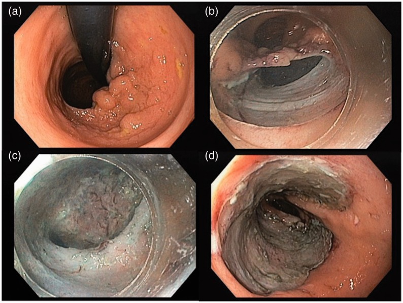
Figure 2.

Endoscopic submucosal dissection of a sigmoid lateral spreading tumour granular nodular mixed type (high-grade dysplasia, en-bloc resection, free margins). a: proximal margin of the lesion in retroflexed position; b: pocket method, with tunnel creation; c: image inside the pocket; d: lesion site after en-bloc ESD.
