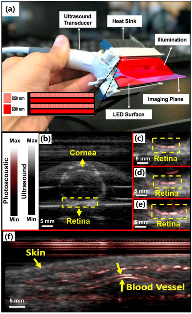
Figure 9.
(a) PLED-PAI probe with imaging plane and illumination source are shown schematically. LED array design is also shown in the inset—there were alternating rows of LEDs with different wavelengths. (b–e) Evaluation of PLED-PAI on rabbit eye. (b) B-mode ultrasound image when fresh enucleated rabbit eye was embedded in 1% agar. (c) B-mode photoacoustic/ultrasound image of rabbit eye using 690 nm (d) B-mode Photoacoustic/ultrasound image of rabbit eye using 850 nm. (e) B-mode photoacoustic/ultrasound image when both 690 and 850 nm are used at the same time. Retinal vessels are imaged in a depth of 2 cm. (f) Photoacoustic image of skin and vasculature. Skin and blood vessel are shown using yellow arrows. Reproduced with permission from Ref. [68].

