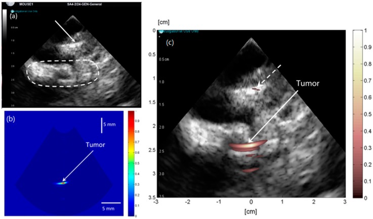
Figure 15.
(a) Pure ultrasound image showing the bottom edge of the plastic seat (arrow) and region of interest (oval); (b) improved post-amplification PA image of tumor in right thigh of nude mouse. (c) Improved filtered PA image superimposed on the pure ultrasound image of the right thigh of the mouse. Reproduced with permission from Ref. [94].

