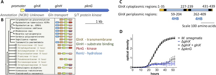
FIG 2 .
PknG is co-expressed with and functionally linked to putative sensor proteins GlnH and GlnX. (A) The PknG operon of M. tuberculosis has a single promoter, and the three genes are co-expressed (49). (B) The structure of the PknG operon is conserved widely in the Actinomycetales (STRING database [42]). (C) The transmembrane segments and membrane topology of GlnX were predicted using TMHMM (38), and putative four-helix bundle sensory domains (4HB) were detected using pfam (39). Putative transmembrane helices are shown in black, cytoplasmic regions in orange, and periplasmic regions in blue. (D) Deletion of glnX from M. smegmatis mirrored the phenotype of pknG-deficient M. smegmatis: a defect in utilization of glutamate as the sole nitrogen source. Growth of glnX-deficient M. smegmatis was restored by reintroduction of glnX. Error bars represent standard deviations from three wells, and the growth curve is representative of three independent experiments.

