
FIG 4 .
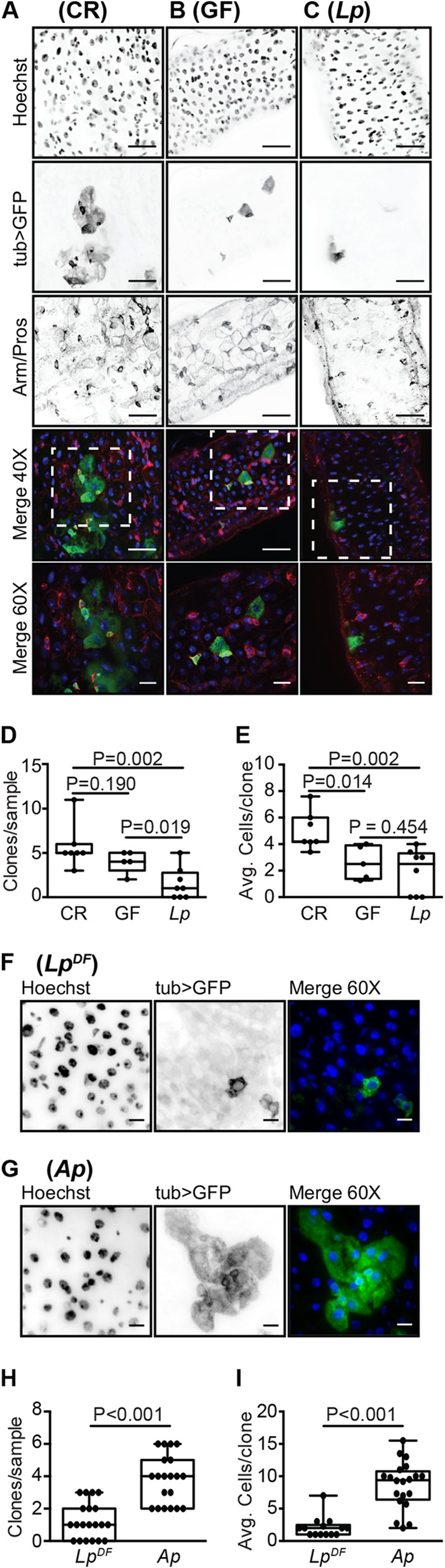
A lack of epithelial renewal in the guts of L. plantarum-monoassociated flies. (A to C) GFP-positive MARCM clones from the posterior midgut of CR (A), GF (B), and L. plantarum-monoassociated (C) flies at day 26 of age. Guts were stained with Hoechst stain and anti-Armadillo/Prospero antibodies as indicated. Hoechst stain (blue), GFP (green), and Armadillo/Prospero (red) were merged in the fourth (×40 magnification) and fifth rows. Boxed regions in the fourth row are shown at a higher magnification (×60) in the fifth row. (D and E) Quantification of clones per sample (D) and cells per clone (E) in CR, GF, and L. plantarum-monoassociated flies. (F and G) GFP-positive MARCM clones from the posterior midgut of L. plantarum DF-monoassociated (F) or A. pasteurianus-monoassociated (G) flies at day 26 of age. Guts were stained with Hoechst stain. Hoechst stain (blue) and GFP (green) were merged in the third column (×60). (H and I) Quantification of clones per sample (H) and cells per clone (I) in L. plantarum DF- and A. pasteurianus-monoassociated flies. For all images, ×40 bars are 25 µm and ×60 bars are 10 µm. P values are the results of pairwise comparisons from a one-way ANOVA.