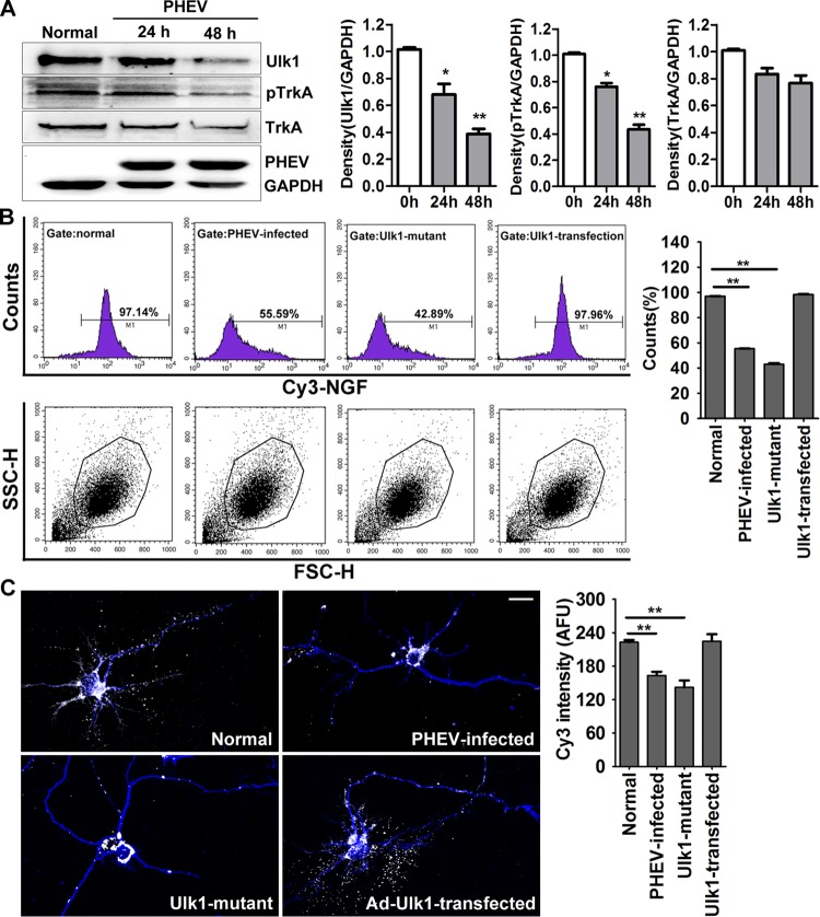
FIG 4.
PHEV infection suppresses the Ulk1-mediated endocytosis of NGF/TrkA complexes. (A) Cortical neurons were incubated with PHEV as indicated, and cell lysates were analyzed by Western blotting with antibodies against Ulk1, TrkA, pTrkA, PHEV, and GAPDH. Ulk1 and pTrkA expression were inhibited by PHEV, although the total expression of TrkA was not significantly affected. Densitometric analysis was performed; Ulk1, pTrkA, and TrkA intensities were normalized against the amount of GAPDH. (B) Cy3-NGF internalization assay: Cy3-NGF in the Ad-GFP-Ulk1-transfected, Ulk1 mutant, PHEV-infected, and normal cortical neurons was detected by FACS, and the data were normalized to internalization rates in normal neurons. Representative FACS profiles obtained during the analysis are shown, and Cy3 signals were gated in M1 and expressed as a percentage of the total number of cells analyzed. SSC-H, side scatter height; FSC-H, forward scatter height. (C) Representative images of Cy3-NGF (white) internalization in the MAP2-marked neurons (blue). Quantitative analyses revealed that the Cy3-NGF internalization was markedly suppressed in the PHEV-infected or Ulk1 mutant neurons. The y axis represents the average Cy3 intensity using arbitrary fluorescence units. Bars, 20 μm. *, P < 0.05; **, P < 0.01 (Student's t test).

