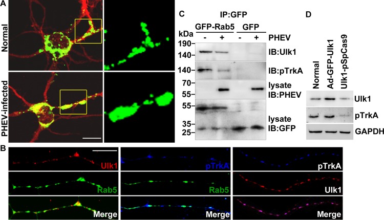
FIG 6.
Ulk1 is associated with TrkA within Rab5 endosomes. (A) Cortical neurons were transfected with GFP-Rab5 at 4 DIV for 24 h and incubated with PHEV at 5 DIV for 24 h and then fixed and subjected to immunocytochemistry with an antibody against MAP2 (red). Typical images of GFP-Rab5-expressing neurons are shown in the left panels. Boxed regions are shown at a higher magnification in the right panels. Bar, 20 μm. (B) Cortical neurons (4 DIV) were transfected with GFP-Rab5, fixed, and subjected to immunocytochemistry with antibodies against Ulk1 (red) and pTrkA (blue) at 6 DIV. The representative micrographs showed that the Ulk1 and pTrkA dots were localized along the growing axon within Rab5 endosomes. Bar, 5 μm. (C) GFP-Rab5- or GFP-expressing neurons were infected with PHEV for 24 h, and the cell lysates were immunoprecipitated (IP) by anti-GFP antibody-conjugated Sepharose beads, followed by SDS-PAGE and immunoblot (IB) analysis with the indicated antibodies. (D) With pretreatment of Ad-GFP-Ulk1 transfection or CRISPR/Cas9-mediated Ulk1 mutant in the cortical neurons, the cell lysates were subjected to Western blot assay to determine the levels of Ulk1 and pTrkA.

