FIG 2.
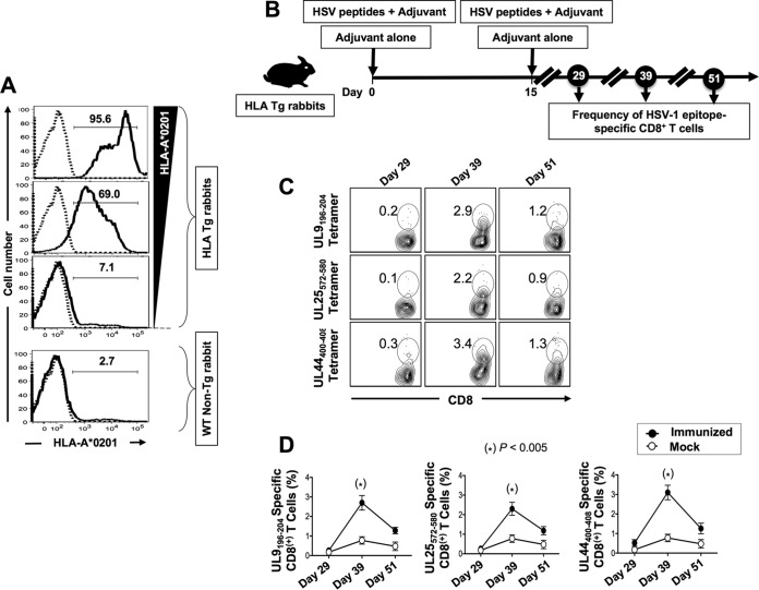
Frequencies of UL9196–204, UL25572–580, and UL44400–408 tetramer-specific CD8+ T cells induced in HLA Tg rabbits following peptide immunization. (A) Detection of HLA-A*02:01 molecules in PBMCs from HLA Tg rabbits. PBMCs from either HLA Tg rabbits or wild-type nontransgenic rabbits were stained first with the BB7.2 MAb and then with a PE-conjugated anti-mouse secondary IgG antibody and analyzed by flow cytometry (see Materials and Methods). (B) Illustration of UL44, UL9, and UL25 peptide immunization regimen and immunological characterization. HLA Tg rabbits were immunized twice, on days 0 and 15, with a mixture of the UL9196–204, UL25572–580, and UL44400–408 peptides in CpG (ODN2007) adjuvant, or they received adjuvant alone (mock group). (C) HLA Tg rabbits were bled on days 29, 39, and 51 after initial immunization (i.e., on days 14, 24, and 36 after the second immunization), and the frequencies of UL9196–204, UL25572–580, and UL44400–408 epitope-specific CD8+ T cells were detected using the corresponding HLA-A*02:01-peptides/tetramers. Shown are representative contour plots of the frequencies of tetramer+ CD8+ T cells specific to the UL9196–204, UL25572–580, and UL44400–408 epitopes detected in PBMCs from immunized rabbits. The number at the top of each box represents the percentage of epitope-specific CD8+ T cells. (D) Average frequencies of tetramer-specific CD8+ T cells detected in all animals on days 29, 39, and 51 after initial immunization. Solid circles represent peptide-immunized rabbits. Open circles represent mock-immunized rabbits. The results are representative of two independent experiments.

