FIG 1.
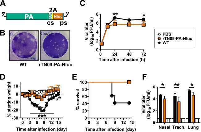
rTN09-PA-Nluc H1N1 influenza virus is attenuated in MDCK cells and mice. (A) Schematic representation of the PA-Nluc cDNA. The codon swap (cs) region, self-cleaving 2A peptide, NanoLuc luciferase (Nluc), and packaging sequence (ps) are indicated. (B) Plaque morphologies of WT and rTN09-PA-Nluc after 2 days of infection in MDCK cells at 37°C. (C) Multistep replication kinetics of WT and rTN09-PA-Nluc in MDCK cells. MDCK cells were infected at an MOI of 0.001, and culture supernatants were collected at the indicated times and titrated by plaque assay in MDCK cells. (D and E) Body weight changes (D) and survival (E) were monitored daily for mice intranasally inoculated with 30 μl PBS containing 750 PFU of WT or rTN09-PA-Nluc. (F) Nasal turbinates, trachea, and lungs were harvested from the mice 4 days postinfection (dpi), and virus titers in tissue homogenates were determined by plaque assay. The reported values are means and standard deviations (n = 5). *, P < 0.05; **, P < 0.01; ***, P < 0.001.

