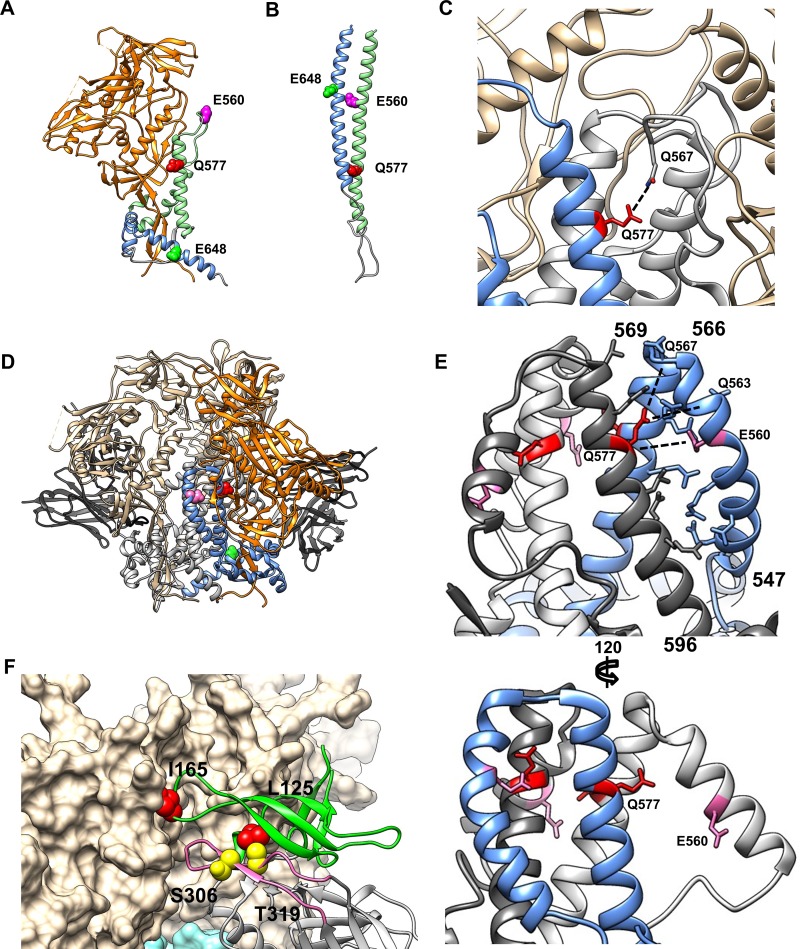
FIG 6.
Model of gp41 and gp120 interactions. (A) gp120-gp41 monomer from the BG505 SOSIP trimer structure (PDB accession number 5CEZ). gp120 is shown in orange, gp41 HR1 is in light green, and gp41 HR2 is in blue. Key gp41 positions E560, Q577, and E648 are shown in magenta, red, and bright green, respectively. (B) gp41 monomer from the postfusion 6HB structure (PDB accession number 1IF3), colored as described above for panel A. (C) Closeup of the N-HR1 region from the SOSIP trimer structure under PDB accession number 5CEZ. One protomer of gp41 is shown in blue, and the others are in gray; gp120 is in beige. Residue Q577 is highlighted in red, and the potential interaction with Q567 from an adjacent gp41 monomer is indicated. (D) JRFL trimer structure (PDB accession number 5FUU), stabilized by two PGT151 Fabs. A single gp120/gp41 protomer is colored as described above for panel A; the remaining gp120/gp41 protomers are shown in beige and gray. PGT151 Fabs are in black. (E) Closeup of the N-HR1 region of gp41 from the structure reported under PDB accession number 5FUU. Each gp41 protomer is colored separately; the unliganded protomer is shown in blue, and the PGT151-liganded protomers are shown in white and charcoal. (Top) Side view of the unliganded (blue) protomer showing ladder-like packing between the N-HR1 and C-HR1 regions. (Bottom) Side view of a PGT151 liganded protomer showing extension of N-HR1 up and away from the C-HR1 coiled coil. (F) Model of key V1/V2 and V3 gp120 mutations in trimer structure. One protomer of gp120 (PDB accession number 4TVP) is shown as a ribbon diagram with colored domains: V1/V2 is in green, the V3 loop is in pink, and the remainder is in gray. The other two gp120 protomers are shown in beige space-fill. Key V1/V2 residues L125 and I165 are highlighted in red. Key V3 residues S306 and T319 are highlighted in yellow.

