Figure 1.

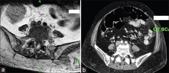
(a) Magnetic resonance imaging and (b) computed tomography images showing compression of the left common iliac vein by the right common iliac artery at its origin against the lumbar spine

(a) Magnetic resonance imaging and (b) computed tomography images showing compression of the left common iliac vein by the right common iliac artery at its origin against the lumbar spine