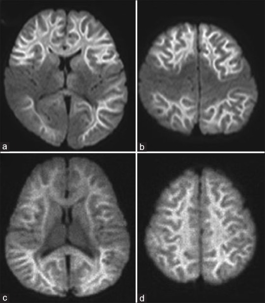
Figure 1.

Diffusion-weighted image of acute leukoencephalopathy with restricted diffusion. (a and b) Central-sparing acute leukoencephalopathy with restricted diffusion shows sparing of the primary sensory motor cortex; (c and d) in diffuse acute leukoencephalopathy with restricted diffusion, there is diffuse bilateral involvement of the white matter. The white matter shows “bright tree appearance,” which represents high-signal intensity on diffusion-weighted image in the widespread subcortical white matter, which has the appearance of tree branches
