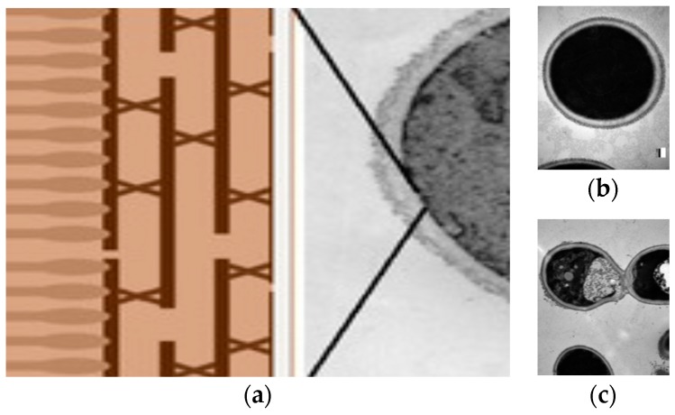
Figure 5.
(a) Schematic molecular structure of yeast cell wall where the inner skeleton shows cross-linked polysaccharides and chitin parallel to the cell surface with an outer dense brush of glycoproteins [104]. (b) Transmission electron micrographs (TEM) of Candida albicans control. (c) TEM of Candida albicans damaged by a cationic antimicrobial polymer showing the burst of the cell membrane [106]. Adapted with permission from [106]. Copyright (2012) American Chemical Society.

