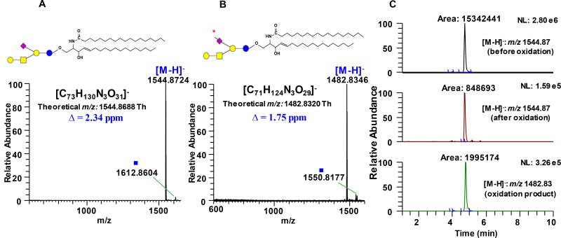
Figure 2.
Full scan MS spectrum of unoxidized (A) and oxidized (B) ganglioside GM1a d18:1/18:0 acquired using LTQ Orbitrap XL demonstrating almost negligible side reactions. (C) Representative extracted ion chromatograms (EIC’s) of GM1a d18:1/18:0 substrate before oxidation (panel 1), residual unoxidized substrate left after incubation with 1mM NaIO4 (panel 2), and the resulting product of oxidation (panel 3). Guide to symbols:
 : aldehyde at C-7 position of Neu5Ac side chain; Δ: absolute mass error; ■: tentatively assigned as sodium formate adduct, [M+HCOO−+Na+-H]− based on accurate mass measurement and isotopic distribution. Structure and systematic name of gangliosides are shown in Supporting Information Table S1. Full scan mass range of other gangliosides subjected to NaIO4 oxidation is shown in Supporting Information Fig. S3 & S4.

