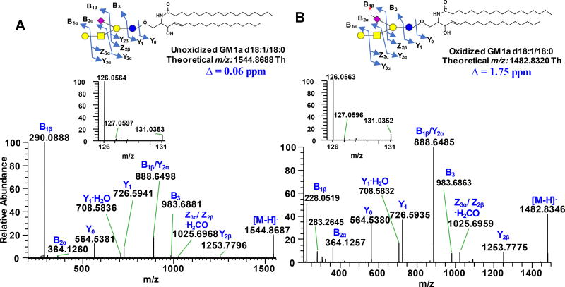
Figure 4.
Negative ESI-MS/MS spectra of unoxidized (A) and oxidized (B) ganglioside GM1a d18:1/18:0 acquired using Q Exactive HF. Asterisk (
 ) on the Neu5Ac symbol indicates aldehyde at C-7 position. Insets show the magnified m/z 126–131 region. Δ indicates absolute mass error. Fragments are labeled according to Domon and Costello nomenclature43. Structure and systematic name of gangliosides are shown in Supporting Information Table S1.

