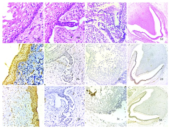
Figure 1.
Staining of stages of odontogenesis: (1a) shows H and E stained dental lamina at x400; (1b) shows H and E stained bud stage at x400; (1c) shows H and E stained cap stage at x400; (1d) shows H and E stained bell stage at x100. (2a) shows BMP4 stained dental lamina at x400, the epithelium is strongly positive with moderate reactivity in ectomesenchyme; (2b) shows BMP4 stained bud stage at x400, the epithelium is nonreactive with slight reactivity in ectomesenchyme; (2c) shows BMP4 stained cap stage at x400, the epithelium and ectomesemchyme are nonreactive; (2d) shows BMP4 stained bell stage at x100, the epithelium and dental papilla is strongly positive. (3a) shows FGF8 stained dental lamina at x400, the epithelium is strongly positive with moderate reactivity in ectomesenchyme; (3b) shows FGF8 stained bud stage at x400, the epithelium and ectomesenchyme are nonreactive; (3c) shows FGF8 stained cap stage at x400, the epithelium and ectomesenchyme are nonreactive; (3d) shows FGF8 stained bell stage at x100, the epithelium and dental papilla is strongly positive.

