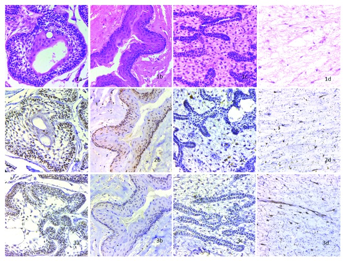
Figure 2.
Staining of odontogenic tumors and odontogenic cyst: (1a) shows H and E stained SMA at x400; (1b) shows H and E stained OKC at x400; (1c) shows H and E stained AF at x400; (1d) shows H and E stained OM at x400. (2a) shows BMP4 stained SMA at x400, the epithelium is positive with slight reactivity in mesenchyme; (2b) shows BMP4 stained OKC at x400, the epithelium is strongly reactive with moderate reactivity in mesenchyme; (2c) shows BMP4 stained AF at x400, the epithelium and mesenchyme are mildly reactive; (2d) shows BMP4 stained OM at x400, the mesenchymal component is strongly positive. (3a) shows FGF8 stained SMA at x400, the epithelium is strongly positive with moderate reactivity in mesenchyme; (3b) shows FGF8 stained OKC at x400, the epithelium is positive and mesenchyme shows slight reactivity; (3c) shows FGF8 stained AF at x400, the epithelium and mesenchyme are mildly reactive; (3d) shows FGF8 stained OM at x400, the mesenchyme is strongly positive.

