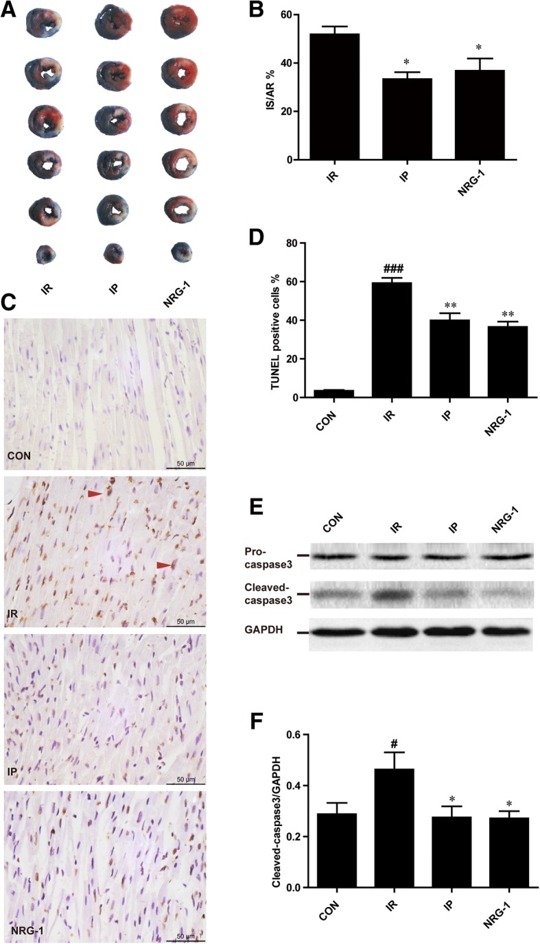
Fig. 1.

Protective effects of both IP and NRG-1 by reducing the IS and apoptosis induced by IR in vivo. (a), Representative heart slices stained by Evans blue and TTC. Blue: non-ischaemic area; non-blue: the area at risk (AR); white: infarct size (IS). (b), The percentage of infarct size/area at risk (IS/AR%). (c), Representative myocardial apoptosis in paraffin sections of the heart at the risk area. The normal cellular nuclei were stained blue by haematoxylin; the apoptotic nuclei were stained brown by TUNEL assay. (d), The percentage of TUNEL-positive cells in the total cells. (e), Representative protein levels of pro-caspase 3 and cleaved-caspase 3 by western blotting. (f), Semi-quantification of cleaved-caspase 3 protein levels normalised to GAPDH. CON: control, IR: ischaemia-reperfusion, IP: ischaemic postconditioning, NRG-1: IR + NRG-1. Data are shown as the mean ± SEM (n = 6). #p < 0.05, ###p < 0.001 vs. CON, *p < 0.05, **p < 0.01 vs. IR
