Fig. 3.
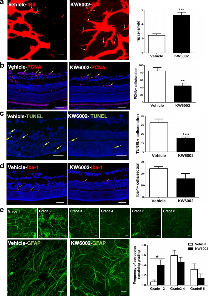
In OIR, the effect of KW6002 treatment on cellular proliferation, tip cell, astrocytes and microglial numbers, and apoptosis in retina at P17. a Endothelial tip cells in retina at P17 of OIR were stained with isolectin B4 (red). Scale bar: 20 μm. Quantitative analysis shows that KW6002 treatment increased tip cell number compared to the vehicle-treated pups (***p < 0.001, Student’s t-test, n = 11–13/group). b Cell proliferation in retina was assessed by immunohistochemistry of PCNA at P17 of OIR. PCNA+ cells (yellow arrow) were quantified. (**p < 0.01, Student’s t-test, n = 8/group). Scale bar: 50 μm. c Apoptotic cells in retina were analyzed by TUNEL (green) staining and individual cells were visualized by DAPI (blue) staining at P17 of OIR. Retinal TUNEL-positive cells (yellow arrow) were quantified and analyzed. KW6002 treatment reduced cellular apoptosis in (**p < 0.01, Mann-Whitney U test, n = 8–9/group). Scale bar: 50 μm. d Microglial activation in retinas was assessed by immunofluorescence staining of Iba-1 at P17 of OIR. Retinal Iba-1-positive cells (yellow arrow) were quantified and analyzed. (p > 0.01, Student’s t-test, n = 8/group). Scale bar: 50 μm. e Representative images show GFAP-positive cells in the avascular areas by anti-GFAP (green) staining at P17 of OIR. Scale bar: 20 μm. GFAP staining in the avascular area was graded on a scale from 1 to 6 as described in the Methods section. The grades of astrocytes from each treatment group were analyzed at P17 of OIR. KW6002 treatment enhanced GFAP staining (with the reduced grade) (*p < 0.05, Mann-Whitney U test, n = 9–12/group), Scale bar: 20 μm

