Fig. 3.

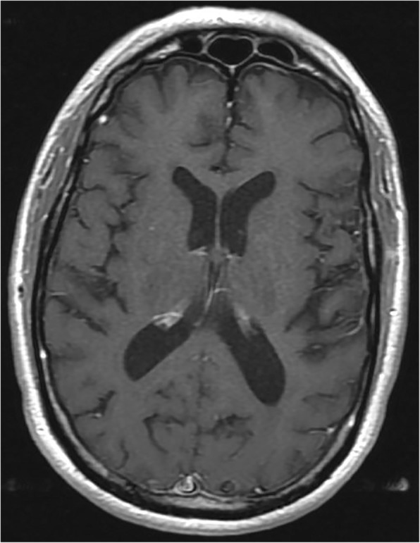
Brain MRI obtained 4 months after starting methotrexate in setting of neurological improvement. T1-weighted contrast-enhanced axial image through the parieto-occipital region shows resolution of leptomeningeal enhancement

Brain MRI obtained 4 months after starting methotrexate in setting of neurological improvement. T1-weighted contrast-enhanced axial image through the parieto-occipital region shows resolution of leptomeningeal enhancement