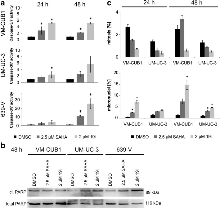
Fig. 2.
Cellular effects of 19i treatment in urothelial carcinoma cell lines. a Caspase 3/7 activity (24 and 48 h) and b cleaved PARP (48 h) were monitored in UCCs VM-CUB1, UM-UC-3, and 639-V after treatment with 19i (2 μM) or SAHA (2.5 μM). c Quantitative analysis of nuclear morphology, based on DAPI stainings, in UCCs VMCUB1 and UM-UC-3. The percentages of mitoses and micronuclei are shown after treatment with HDACi 19i (2 μM), SAHA (2.5 μM), or DMSO for 24 or 48 h. The calculated significances refer to the DMSO solvent control (*p < 0.05). Data in a and c are mean from n = 3; the blot in b shows a representative experiment

