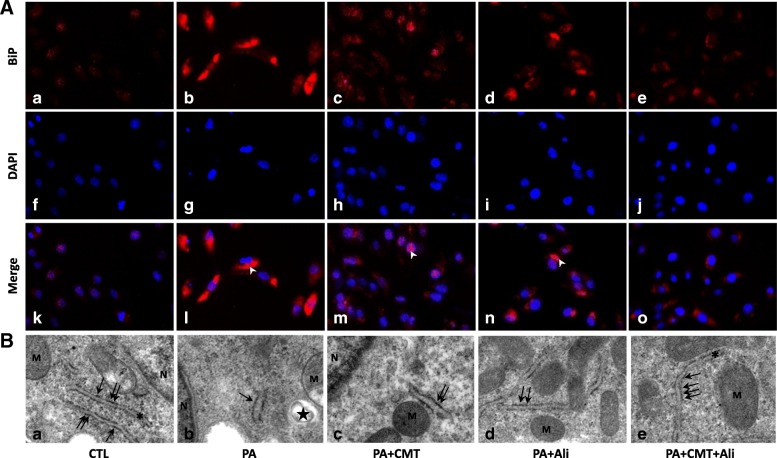
Fig. 2.
Combination treatment with chymostatin and aliskiren attenuated ER stresss in HK2 cells treated with PA. a Immunofluorescence staining of BiP in cultured HK2 cells. In controls (a, f, k), BiP scarcely localizes intracellular cytoplasm of HK2 cells, whereas PA (0.6 mM) induced significantly increased labeling of BiP (b, g, l), which was suppressed by either chymostatin (5X10⁻⁵M) (c, h, m), aliskiren (10− 8 M) (d, i, n) or their combination treatment (e, j, o). b Representative images of endoplasmic reticulum morphology by transmission electron microscopy in HK2 cells. (a) controls; (b) PA treatment group; (c) PA plus chymostatin treatment; (d) PA plus aliskiren treatment; (e) PA plus chymostatin and aliskiren treatment. N: nucleus; M: mitochondria; asteroids: endoplasmic reticulum; the star: lipid drop; arrows: ribosome; Magnification 37,000× CTL, controls; PA, palmitic acid treatment group; PA + CMT, palmitic acid plus chymostatin treatment; PA + Ali, palmitic acid plus aliskiren treatment; PA + CMT + Ali, palmitic acid plus chymostatin and aliskiren treatment; arrow heads: BiP; asterisks: ER; M: mitochodria; N: nucleus

