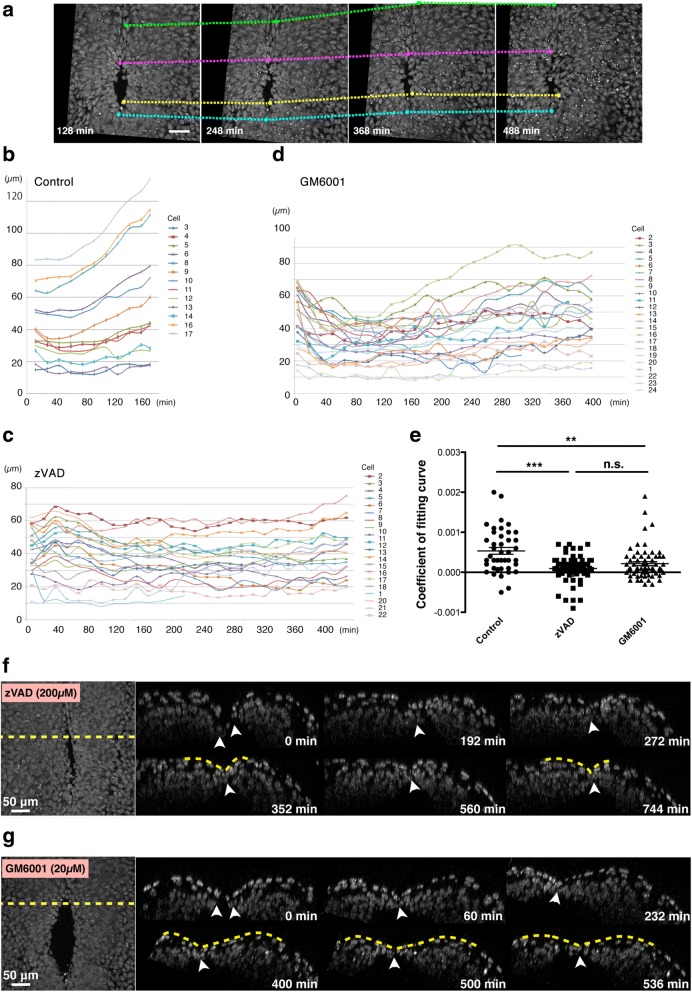
Fig. 4.
Inhibition of matrix metalloproteases (MMPs) prevents backward movement. a Time-lapse images of the mid-hindbrain neuropore (MHNP) closure in the presence of an inhibitor of MMPs, GM6001. b, c, d Examples of tracking of cells on the neural ridge during the neuropore closure of control (b), Z-VAD-FMK-treated (c), and GM6001-treated (d) embryos. Cells on the neural ridge were chosen evenly along the circumference of the neuropores, and their distance to a randomly-selected standard cell (Y-axis) was measured and plotted at each time-points (X-axis). Plots were created from every 16-min time-point, although images used for the tracking analysis were acquired every 4 min. Three embryos per each condition were processed for this analysis, and representative ones were presented. e Comparison of the speed of backward movements of cells on the neural ridge by squared term coefficients of fitting curves. Treatments with Z-VAD-FMK and GM6001 significantly reduced the speed of the backward movement (**p < 0.01, ***p < 0.001, one-way ANOVA). Each dot indicates one cell, and 41, 64, and 66 cells from three embryos per conditions were analyzed for control, Z-VAD-FMK, and GM6001, respectively. f, g Time-lapse montages of transverse (xz) sections reconstituted form 4D (x, y, z, t) dataset during the MHNP closure of Z-VAD-FMK-treated (f) and GM6001-treated (g) embryos. A dotted line in left panel (xy images) indicates the position for making xz sections shown in right panels. Scale bars: 50 μm

