Figure 1.

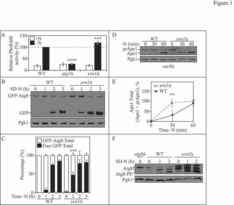
Xrn1 negatively regulates autophagy. (A) WT (WLY176), atg1∆ (WLY192) and xrn1∆ (EDA62) cells were grown to mid-log phase in YPD (+N) and then starved for nitrogen (-N) for 3 h. The Pho8Δ60 activity was measured and normalized to the activity of starved WT cells, which was set at 100%. Results are the mean of 5 independent experiments. Error bars indicate standard deviation (SD). (B) WT (WLY176) and xrn1∆ (EDA62) cells transformed with a centromeric plasmid encoding GFP-Atg8 were grown to mid-log phase in rich selective medium and then starved for nitrogen (SD-N) for 0, 1, 2 or 3 h. Cells were collected at the indicated time points and protein extracts were analyzed by SDS-PAGE and blotted with anti-YFP or anti-Pgk1 (loading control) antibodies. (C) Densitometry of blots represented in (B). The ratio of GFP-Atg8:total GFP (sum of full-length GFP-Atg8 and free GFP) or free GFP:total GFP was quantified with ImageJ. Results shown are the mean of 4 independent experiments +/− SD. (D) WT (vac8∆; CWY230) and xrn1∆ (vac8∆ xrn1∆; EDA152) cells at 0, 30 and 60 min of nitrogen starvation. Precursor (prApe1) and mature (Ape1) forms of aminopeptidase 1 were detected with anti-Ape1 or anti-Pgk1 (loading control) antisera. (E) Densitometry of blots represented in (D). The ratio of Ape1:total Ape1 (sum of prApe1 and Ape1) was quantified with ImageJ. Results shown are the mean of 4 independent experiments +/− SD. (F) WT (SEY6210), atg8∆ (YAB369) and xrn1∆ (EDA85) cells were grown to mid-log phase in YPD and then starved for nitrogen (SD-N) for 0, 1, or 2 h. Protein extracts were analyzed by SDS-PAGE and blotted with anti-Atg8 or anti-Pgk1 (loading control) antisera (*denotes nonspecific band). Blots shown are representative of 3 independent experiments. Also see Table S1.
