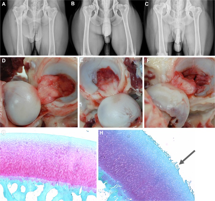
Figure 1.
Anatomy of canine hip dysplasia.
Notes: (A–C) Canine hip-extended radiographs, and corresponding images of the joints (D–F) from different individuals demonstrating mild (A and D), moderate (B and E), and severe (C and F) joint changes. Light photomicrographs of normal (G) and fibrillated (H, arrow) articular cartilage.

