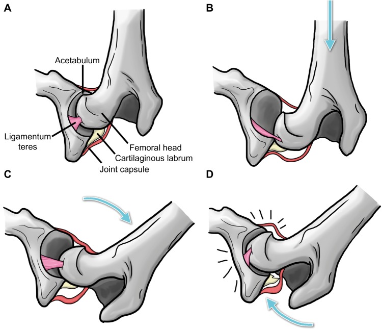
Figure 2.
Schematic illustration of the Ortolani test.
Notes: Image demonstrates the coxofemoral joint prior to distraction (A), while force is applied from the stifle toward the hip along the axis of the femur to displace the femoral head (B), during abduction of the femur to reduce the joint (C), and with the femoral head snapping back into place with an audible click, ie, the Ortolani sign (D). Arrows indicate the direction of the applied force. Adapted from Chalman JA, Butler HC. Coxofemoral joint laxity and the Ortolani sign. Journal of American Animal Hospital Association. 1985;21:671–676.28

