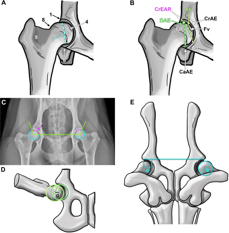
Figure 3.
Representations of anatomical landmarks and evaluation mechanisms to assess canine hip dysplasia.
Notes: Coxofemoral joint anatomical characteristics considered by the Orthopedic Foundation for Animals (A): craniolateral acetabular rim (1), cranial acetabular margin (2), femoral head (3), fovea capitis (4), acetabular notch (5), caudal acetabular margin (6), dorsal acetabular margin (7), junction of femoral head and neck (8), and trochanteric fossa (9). (B) British Veterinary Association/Kennel Club canine coxofemoral joint characteristics scored during evaluation.10,34 Schematic superimposed on a hip-extended radiograph demonstrating the Norberg angle (C, arrow). Illustration of the Pennsylvania Hip Improvement Program (distraction index, the distance between the centers of the femoral head and acetabulum during distraction (D) divided by the radius (r) of the femoral head (d).41 Depiction of the dorsolateral subluxation score (E) calculated as 100 multiplied by the percentage of femoral head medial to the cranial acetabular rim (d) divided by the femoral head diameter (θ), d/θ ×100%).
Abbreviations: AF, acetabular fossa; An, acetabular notch; CaAE, caudal acetabular edge; CrAE, cranial acetabular edge; CrEAR, cranial effective acetabular rim; DAE, dorsal acetabular edge; Fh, femoral head; Fv, foveal defect.

