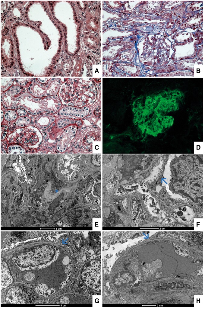
Fig. 2.
Light and immunofluorescence microscopy. (A) Hematoxylin and eosin, magnification × 40, observing focal tubular dilatation. (B and C) Masson's trichrome and periodic acid–Schiff stains, magnification × 40, showing a thin glomerular basement membrane with mild increasing of the mesangial matrix and cellularity. (D) Direct immunofluorescence for immunoglobulin M presenting diffuse mesangial staining. Electron microscopy. (E) Discrete increasing of the mesangial matrix (see asterisk). (F, G and H) Diffuse and severe foot process effacement (see arrows).

