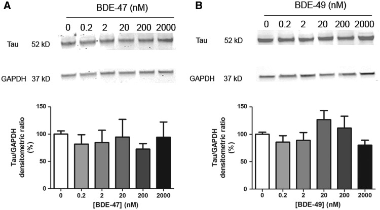
FIG. 4.
BDE-47 and BDE-49 do not decrease tau-1 protein in mature cultures. At DIV 7, hippocampal neurons were exposed to BDE-47 (A) or BDE-49 (B) for 48 h. At the end of the exposure, cells were lysed for western blotting and probed with antibodies specific for tau-1 (axonal cytoskeletal protein) and GAPDH (loading control) as shown in representative western blots (top panels). Bar graphs (bottom panel) represent densitometric data. Densitometric values of tau-1 immunopositive bands were normalized to densitometric values for GAPDH immunopositive bands within the same sample. Data presented as mean ± SE (n = 3 independent dissections). ***P < .001 relative to vehicle control.

