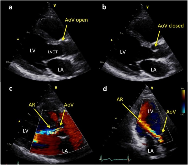
Figure 1.
Echocardiographic images of a thickened and mildly restricted aortic valve (AoV) with resultant moderately severe aortic regurgitation (AR). (a) Parasternal long axis views in which the AoV leaflets are mildly thickened and show mild doming owing to incomplete opening in systole. (b) The AoV leaflet tips are mildly thickened when the valve is closed. (c) Parasternal long axis view showing color Doppler image of a broad jet of AR almost filling the left ventricular outflow tract (LVOT). (d) Apical long axis view of the color Doppler jet of AR reaching the left ventricular apex. LA, left atrium; LV, left ventricle.

