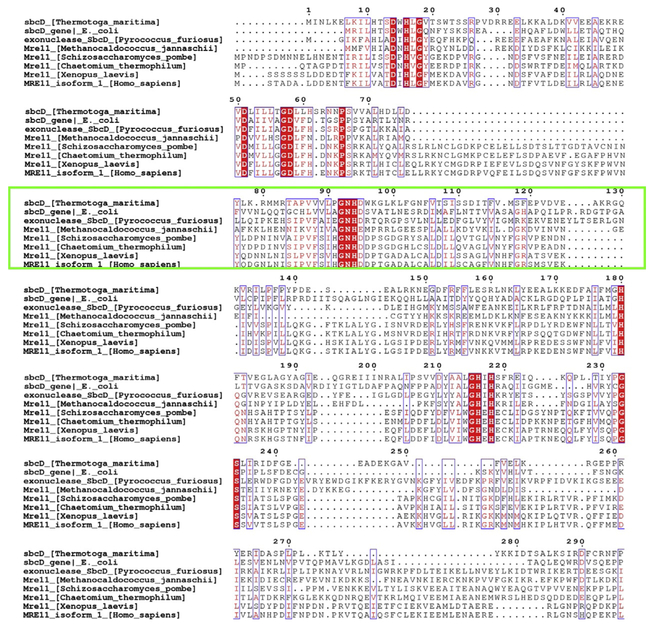
Fig. 2.
Sequence conservation from an alignment performed with Cobalt and Espript 3.0 showing the Mre11 catalytic domain among organisms having a structure deposited into the PDB plus the X. laevis Mre11 used for in vivo identification of Mirin activity. Conserved regions (red fill) and the region showing an allosteric conformational change in T. maritima centered at N93–H94 is highlighted (green box). Organisms from the top are shown like bacterial, through archeal, yeast, fungi, vertebrate, and mammals.

