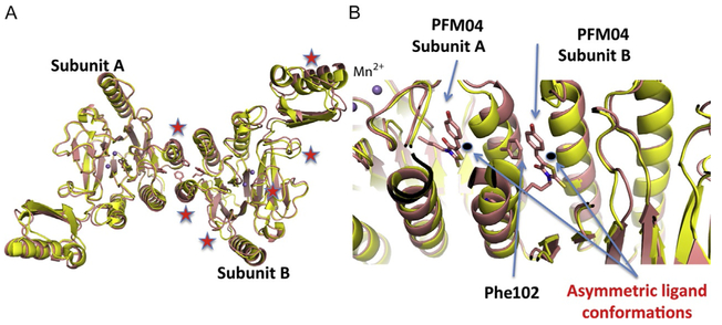
Fig. 5.
The apo and novel PFM04 inhibitor-bound structures. (A) The superimposition of TmMre11 apo (yellow) and the novel TmMre11–PFM04 inhibitor complex structure (pink) showing the different organization of the two domains due to the asymmetric binding of PFM04. Red stars highlight main chain shifts in the inhibited vs the apo structure. (B) A zoom-in on the inhibitor-binding site shows the different conformation of ligands between subunit A and subunit B, along with the interaction of Phe102 in subunit A and PFM04 in subunit B.

