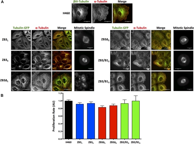
Figure 2. Modification of the βIII-tubulin C-terminal tail does not affect the incorporation of the protein into the microtubule network, the microtubule architecture, or the cellular proliferation rate.
(A) Representative immunofluorescence and live-cell images of the microtubule architecture in gene-edited NCI-H460 cell clones expressing the full-length βIII-tubulin protein (ZB3), truncated βIII-tubulin protein (ZB3Δ), or βIII-tubulin body with the βI-tubulin tail (ZB3/CB1) compared with the NCI-H460 parental cell line (H460). For interphase microtubule architecture, immunofluorescence staining was performed for α-tubulin detection. Individual channels are presented as grayscale images and the merged image with each channel colored accordingly. Left panel: modified βIII-tubulin proteins; center panel: α-tubulin; right panel: merged image of modified βIII-tubulin proteins (green) and α-tubulin (red). Scale bar 25 μm; and far right panel: higher magnification images of the mitotic spindle in live cells (GFP imaging). Scale bar 10 μm. Representative of three independent experiments. (B) Proliferation rates of gene-edited clones as measured by the BrdU assay and normalized to cell number. Mean ± SEM of four independent experiments, no significant difference.

