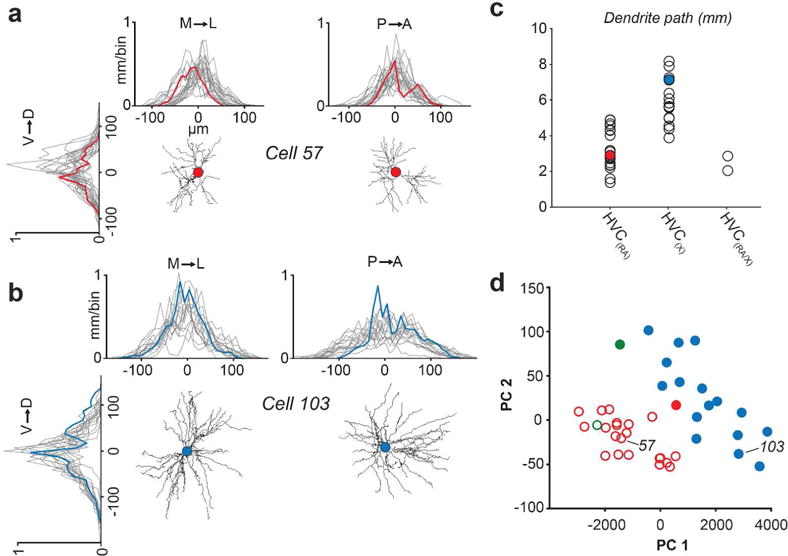
FIGURE 4. Spatial properties of HVC(RA) and HVC(X) dendrites.

(a, b) One dimensional profile of dendritic length along three axes for HVC(RA) (a) and HVC(X) (b) neurons. Red and blue traces are from example neurons, grey traces are from all remaining individual neurons from each group in our data set. The center of each plot (0 μm) denotes soma location. (c) Total dendritic path length organized by projection target. Filled circles represent examples from (a) and (b). (d) The first two principal components of four spatial parameters of dendrites. The two clusters as designated using the OPTICS algorithm (see Materials and Methods) are represented by closed circles and open circles; Red - HVC(RA); Blue - HVC(X); Green - HVC(RA/X).
