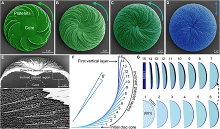
Fig. 4. Platelet layer-by-layer inclination decreases vaterite helicoid chirality toward an achiral, vertical platelet organization.

(A to C) SEM images of the gradual decrease in chirality (green, pseudocolored at 8, 16, and 32 hours of growth) of vaterite helicoids grown in the presence of l-Asp. The helicoids are composed of inclined platelets originating from an initial achiral substrate disc core, growing to the point (note decreasing magnification) where the helicoid is symmetric and achiral [blue in (D), at 72 hours) with vertically oriented platelets at the helicoid surface, and with an increase in overall helicoid size. (E) SEM image of a cross section of a small intermediate counterclockwise chiral vaterite helicoid after FIB cutting, whose interior is composed of two regions—an initial core disc/dome and an inclined-platelet region (top panel). In the inclined-platelet region, the layer-by-layer growth produces inclined-platelet organization having an angle of 6° between a consequential daughter platelet layer (white dashed line on the daughter platelet) arising from a mother platelet layer (white solid line on the mother platelet), which leads to the decrease and disappearance of chirality of the vaterite helicoid as the platelets ascend to the vertical orientation. (F) Side view schematic of growth in the layer-by-layer inclination model where 6° inclination of 15 continuously forming platelet layers arising from an initially flat achiral core disc ascend to the first vertical platelet layer (15th dark blue line). (G) The exposed crystalline vaterite face changes from being initially the basal (001) vaterite plane (light blue) to the (100) plane (dark blue) after the formation of 15 successional nanostructured vaterite platelet layers, with all platelets formed from subunit tilted nanohexagons (19).
