. 2018 Aug 1;7:e33051. doi: 10.7554/eLife.33051

Figure 1. Design and surgical implantation of titanium CLNW.

(A) CAD schematic detailing the dimensions of the titanium plates opposed to fit a chronic lymph node window (CLNW). (B) Following shaving the surgical site, window plates are briefly overlaid for surgical planning and prior to sterile preparation. (C) Following surgical implantation of the CLNW, transillumination confirms the node centered appropriately within the imaging range. (D) Photomicrograph of completed CLNW demonstrating coverglass, incision closure, and LN placement within the center of viewing range. (E) Imaging setup demonstrating anesthetized mouse on a heated H2O reservoir with clamp fixation of the window minimizing motion artifact.

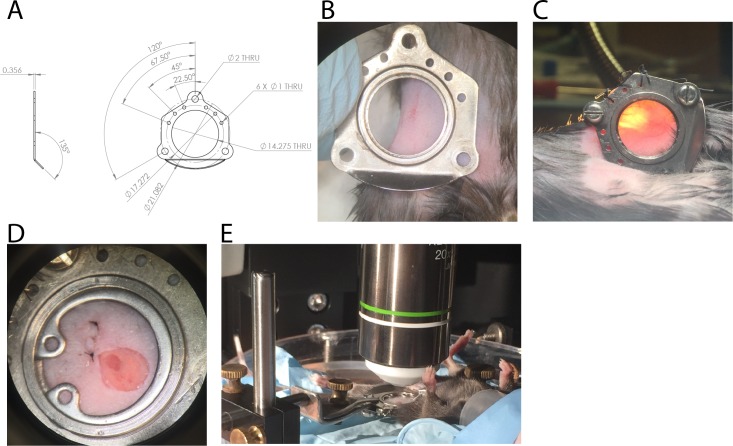
Figure 1.

Figure 1—figure supplement 1. Additional surgical images demonstrates anchor placement and node exposure.

Figure 1—figure supplement 1.

(A) Additional representative image illustrating surgical step just after placing CLNW anchors and prior to stretching skin across the plate and suturing skin anchors. Note LN is visible as darker ovoid shape surrounded by fat, marked by arrowhead. (B) Additional representative image illustrating surgical step after placing and extending the skin incision to include the LN (which is fully encased in the fat pad connective tissue, note sheen in image reflecting this). (C) Additional representative image illustrating surgical step after opening the fat pad capsule and lifting the connective tissue from the superior-most aspect of the LN capsule. Also note, closing a portion of the incision to reduce open surface area. This is prior to sterile irrigation and placement of coverslip.