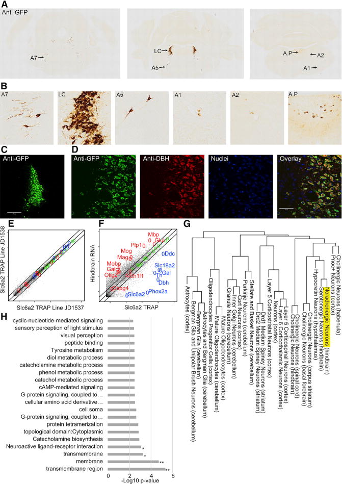
Figure 1. Characterization of Noradrenergic bacTRAP Lines.

(A) Anti-GFP immunohistochemistry demonstrates EGFP/Rpl10a labeling in the hindbrain.
(B) Anti-GFP staining is most robust in anterior groups (A4–A7), especially LC.
(C) Immunofluorescence for GFP (green) labels entire LC. Scale bar, 200 mM.
(D) GFP co-labels completely with dopamine-β-hydroxylase (DBH) (red). Scale bar, 50 mM.
(E) Comparison of two Slc6a2 TRAP lines demonstrates reproducibility.
(F) Slc6a2 TRAP mRNA versus total hindbrain mRNA enriches NE neuron markers (blue) and depletes unrelated cell-type (glial) markers (red). (E and F) Lines indicate 0.5-, 1-, and 2-fold. Log10 scale.
(G) Hierarchical clustering of Slc6a2 neurons.
(H) Slc6a2 TRAP enriches for transmembrane proteins and receptors. Hypergeometric test, Benjamini-Hochberg corrected; *p < 0.06; **p < 0.01.
See also Table S1B.
