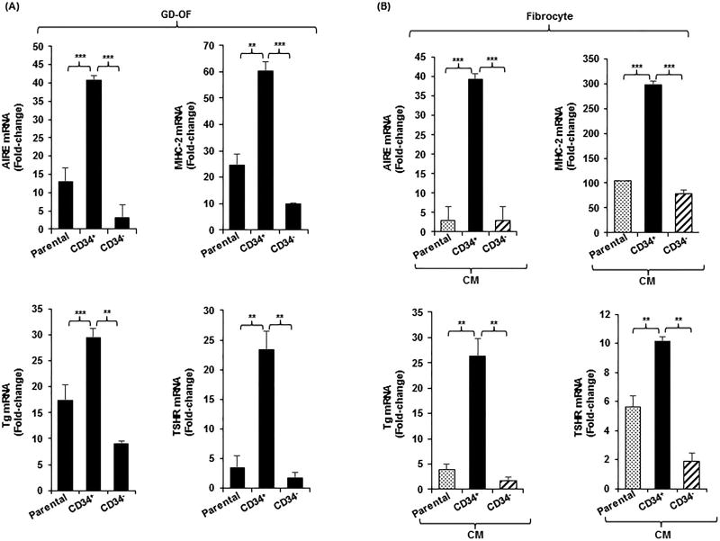
Figure 2.
(A) Expression pattern of AIRE, MHC-2, Tg, and TSHR in parental (mixed) GD-OF and pure CD34+ OF and CD34− OF subsets. Parental GD-OF strains were subjected to sham cytometric sorting or sorted into CD34+ OF and CD34− OF subsets and cultured for 3 d. (B) Conditioned media were generated by incubating GD-OF
 , CD34+ OF
 , and CD34− OF
 in DMEM for 48 h. These media were then used to cover fibrocytes for 3 d. Monolayers were harvested, RNA extracted, reverse-transcribed and subjected to real-time PCR. Data are expressed as the mean ± SD of 3 independent replicates. Values were normalized to their respective GAPDH levels and are expressed as mean ± SD of triplicate determinations. *** p<0.001; ** p<0.01. Experiments were performed three times.

