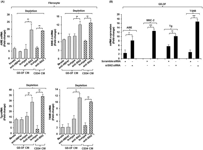
Figure 3.
(A) Depleting medium conditioned by GD-OF of Slit2 restores fibrocyte gene expression. (A) Conditioned media (CM) from GD-OF
 and CD34− OF
 were incubated with uncoated beads or beads coated with anti-Slit2 (100 µg), anti-HSP47 (100 µg), or isotype IgG (100 µg) as described in Methods. Fibrocyte monolayers were incubated with these media for 5 d. (B) Knocking down Slit2 with specific siRNA enhances gene expression in GD-OF. GD-OF were transfected with either scrambled (control) siRNA (3µg) or Slit2-specific siRNA (3µg) and incubated 3 d. RNA was extracted, reversed transcribed, and cDNAs subjected to real-time PCR for the targets indicated. Values were normalized to their respective GAPDH levels and expressed as mean ± SD of triplicate determinations. *** p<0.001, ** p<0.01; * p<0.05. Experiments were performed three times.

