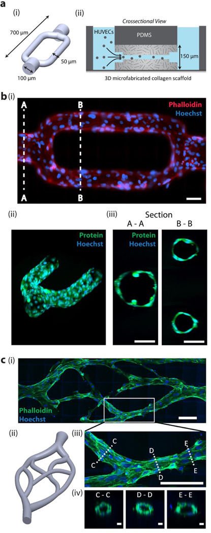
Figure 4. Multi-photon microfabricated microvasculature.
(a) (i) 3D bifurcating microchannel design. The shape represents the unexposed regions in a block of collagen. (ii) Microchannels are microfabricated in a block of collagen within a microfluidic chip to enable addition of HUVECs via hydrostatic pressure driven flow. (b) (i) HUVECs fixed and stained at 12 h in vitro and imaged under (i) epifluorescence, and (ii-iii) 2-photon microscopy. Scale bars = 50 µm. (c) (i-ii) Biomimetic microvascular channels lined with HUVECs, fixed and phalloidin stained at 72 h in vitro. Image is acquired using two-photon microscopy and displayed as a maximum intensity projection. Scale bar = 100 µm. (iii) A maximum intensity image of a bifurcation. Scale bar = 100 µm, and (iv) crossections taken at three points along the vessel, showing a patent lumen. Scale bars = 10 µm.

