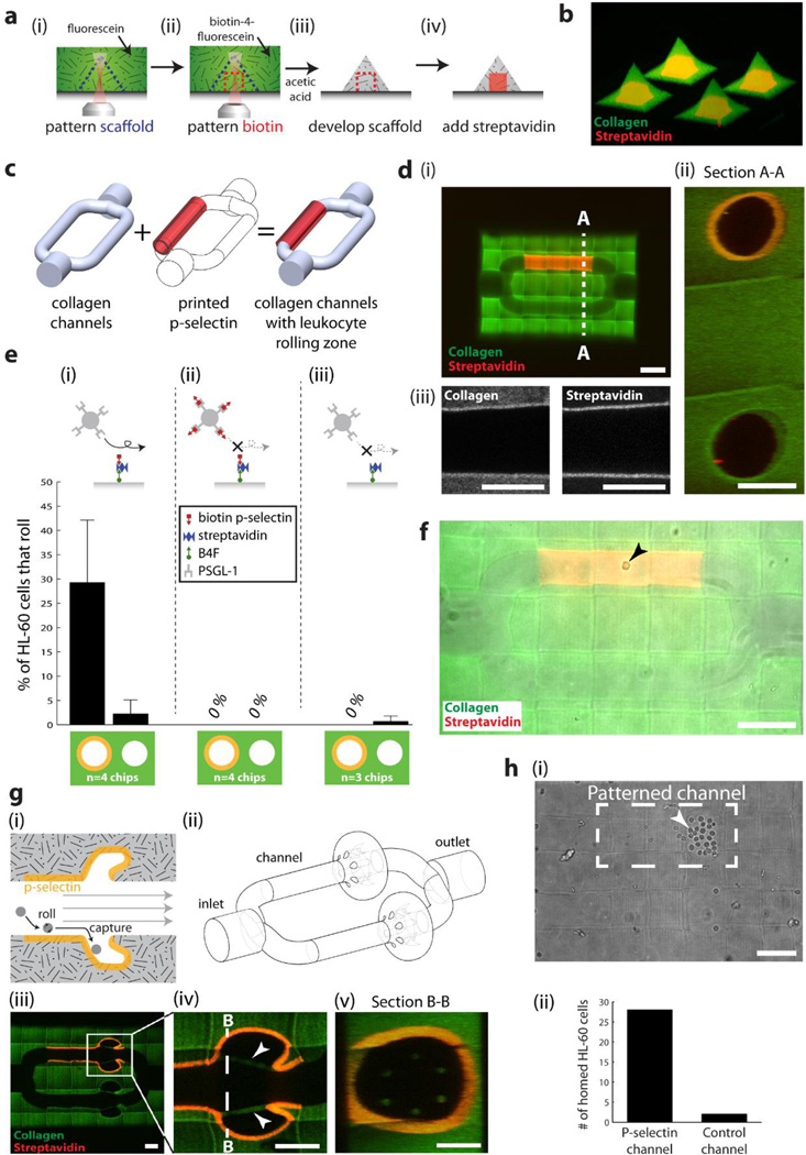
Figure 5. Simultaneous microfabrication of collagen scaffold microarchitecture and patterning of internal protein cues.
(a) Method for microfabricating scaffold microarchitecture and internal patterns of proteins. (b) Microfabricated collagen shapes containing internal patterns of streptavidin. (c) Schematic showing the combination of collagen channels and patterned P-selectin. (d) Collagen channels containing a region patterned with streptavidin (i) Scale bar = 100 µm, (ii) Crossection of channels acquired from two-photon imaging. Scale bar = 50 µm. (iii) Two-photon images of microfabricated collagen and patterned streptavidin. Scale bar = 50 µm. (e) Percentage of HL-60 cells rolling on (i) P-selectin patterned, and non-patterned channels; (ii) the same channels, but the HL-60 cells are pre-incubated with soluble P-selectin; (iii) patterned channels coated with streptavidin only. (f) HL-60 cell rolling on patterned P-selectin (arrowhead). Scale bar = 100 µm. (g) (i) A device to selectively capture rolling cells; (ii) its corresponding 3D model; (iii-v) as microfabricated and imaged using two-photon microscopy. Arrowheads display collagen ‘tethers’ used to support overhanging collagen, and to prevent the overhangs from inverting in conditions of flow. Scale bars = 50 µm. (h) HL-60 cells preferentially home to the side patterned with P-selectin. Scale bar = 100 µm.

