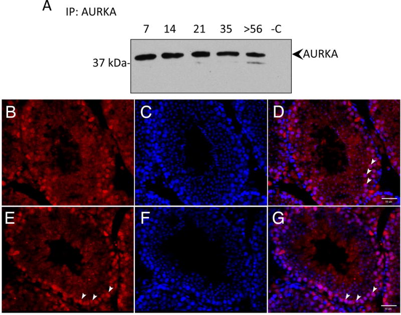
Figure 1. Aurora A kinase is expressed in multiple male germ cell types in the testis.

(A) Protein lysates were prepared from testes of postnatal day 7, 14, 21, 35 and adult mice (>56 dpp). AURKA was precipitated with an AURKA specific antibody and detected with the same antibody. Proteins in lane -C where precipitated in the absence of antibody as control. AURKA expression was detected during the first wave of spermatogenesis in the testis when specific cell types appear at defined times after birth. All cell types can be detected in the adult seminiferous tubule. (B-G) Frozen testes sections were stained with AURKA (red in B and E) and DNA with DAPI (blue in C and F) with the merged image in D and G. (B-D) AURKA localized to spermatogonia (representative spermatogonia indicated with arrowheads). (E-G) In other tubules, AURA localized to spermatocytes (representative spermatocytes indicated with arrowheads). Scale bars indicate 10 μm.
