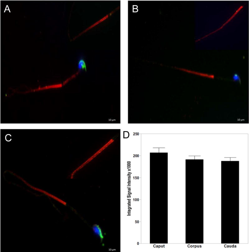
Figure 3. AURKA localizes to the flagellum of epididymal sperm.

Sperm collected from the caput (A), corpus (B) and cauda (C) were triple stained with antibodies to AURKA (red), conjugated PNA-Lectin AF488 (green) for the acrosome and DAPI to visualize the nuclei (blue). AURKA is concentrated at the annulus and expressed throughout the principle piece of the flagellum as two parallel stripes (indicated by arrows in A inset). AURKA was not observed associated with the nucleus, acrosome or midpiece. AURKA expression decreased as sperm traveled through the epididymis, which is graphically represented in (D). Scale bars indicate 10μM.
