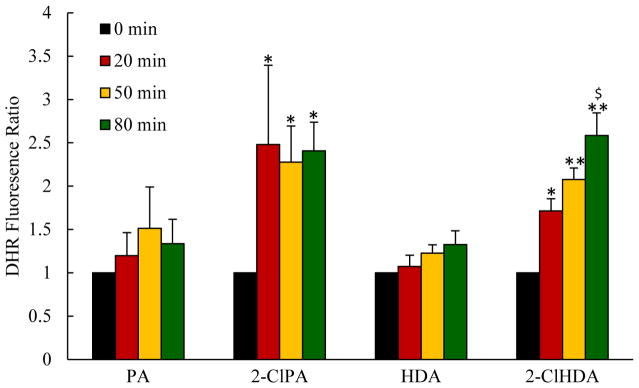
Figure 5.
Mesenteric superfusion with chlorinated lipids increased ROS production in postcapillary venules, as assessed by DHR fluorescence. For each group (n=6), at each time point for every animal, 10 mesenteric venules were randomly chosen and 5 regions of interest (25 um in diameter) along venule wall were measured, averaged and presented relative to baseline (just prior to lipid superfusion). *P<0.05 and **P<0.01 when compared to baseline, $P<0.05 between 2-ClPA and PA, 2-ClHDA and HDA groups at indicated time point.

